Modeling chromosomes with the tasmanian devil answers – Delving into the fascinating realm of modeling chromosomes with the Tasmanian devil, we embark on a journey to unravel the complexities of this enigmatic species’ genetic makeup. The Tasmanian devil’s unique chromosome structure, shaped by evolutionary forces, provides a captivating lens through which we can explore the intricate mechanisms underlying chromosome evolution and its implications for genetic diseases and conservation efforts.
Tasmanian Devil Chromosome Structure: Modeling Chromosomes With The Tasmanian Devil Answers

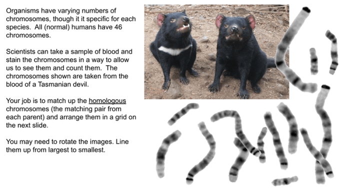
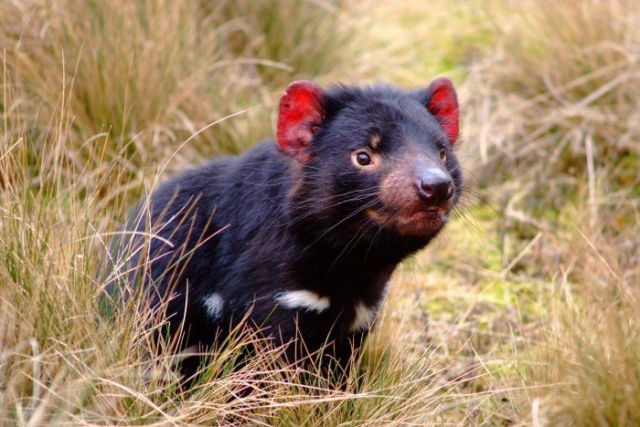
The Tasmanian devil ( Sarcophilus harrisii) is a marsupial species native to the island state of Tasmania, Australia. It is known for its distinctive black fur, white facial markings, and powerful jaws. The Tasmanian devil has a unique chromosome structure that sets it apart from other marsupials.
Tasmanian devil chromosomes are characterized by their large size and high number of telomeres. Telomeres are repetitive DNA sequences that cap the ends of chromosomes and protect them from degradation. The Tasmanian devil has an unusually high number of telomeres, which may be related to its high metabolic rate and short lifespan.
The centromeres of Tasmanian devil chromosomes are also unusual. Centromeres are the regions of chromosomes where spindle fibers attach during cell division. In most mammals, centromeres are located near the center of the chromosome. However, in the Tasmanian devil, centromeres are located near the ends of the chromosomes.
The unique chromosome structure of the Tasmanian devil is thought to be a result of its evolutionary history. The Tasmanian devil is the last surviving member of its genus, and it is believed to have diverged from other marsupials around 10 million years ago.
During this time, the Tasmanian devil’s chromosomes may have undergone significant changes due to genetic drift and natural selection.
Chromosome Modeling Techniques

Various methods can be used to model Tasmanian devil chromosomes. These methods include:
- Fluorescence in situ hybridization (FISH): FISH is a technique that uses fluorescent probes to visualize specific DNA sequences on chromosomes. FISH can be used to identify the location of genes, telomeres, and other chromosomal features.
- Comparative genomic hybridization (CGH): CGH is a technique that compares the DNA content of two different samples. CGH can be used to identify chromosomal rearrangements, such as deletions, duplications, and translocations.
- Microarray analysis: Microarray analysis is a technique that uses DNA probes to measure the expression of genes. Microarray analysis can be used to identify genes that are differentially expressed in different cell types or under different conditions.
Each of these techniques has its own advantages and disadvantages. FISH is a relatively simple and inexpensive technique, but it can only be used to visualize a limited number of DNA sequences. CGH is a more powerful technique, but it is also more expensive and time-consuming.
Microarray analysis is a versatile technique that can be used to measure the expression of thousands of genes, but it is also the most expensive and time-consuming technique.
Modeling Chromosome Evolution

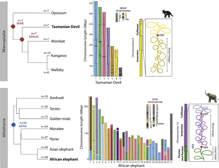
Chromosome modeling can be used to study the evolutionary history of chromosomes. By comparing the chromosome structure of different species, researchers can identify chromosomal changes that have occurred over time. These changes can provide insights into the evolutionary relationships between species and the forces that have shaped their genomes.
The chromosome structure of the Tasmanian devil has been studied extensively to understand its evolutionary history. Researchers have found that the Tasmanian devil has a high rate of chromosomal rearrangements, which may be due to its small population size and high metabolic rate.
These rearrangements have likely played a role in the evolution of the Tasmanian devil’s unique chromosome structure.
Chromosome modeling can also be used to study the role of genetic drift and natural selection in shaping chromosome structure. Genetic drift is the random change in gene frequencies that occurs in small populations. Natural selection is the process by which individuals with favorable traits are more likely to survive and reproduce.
Genetic drift and natural selection can both lead to changes in chromosome structure. Genetic drift can cause the loss of beneficial alleles and the accumulation of harmful alleles. Natural selection can favor chromosomal rearrangements that provide a selective advantage. By studying the chromosome structure of different populations, researchers can identify the role of genetic drift and natural selection in shaping chromosome evolution.
Chromosome Modeling Applications

Chromosome modeling can be used to study a variety of genetic diseases and conservation efforts. For example, chromosome modeling has been used to identify chromosomal rearrangements that cause genetic diseases such as Down syndrome and cystic fibrosis. Chromosome modeling has also been used to track the genetic diversity of endangered species and to identify populations that are at risk of extinction.
One of the most successful applications of chromosome modeling has been in the conservation of the Tasmanian devil. The Tasmanian devil is a critically endangered species that is threatened by a contagious cancer known as devil facial tumor disease (DFTD).
DFTD is a transmissible cancer that spreads through direct contact between devils. It causes large, disfiguring tumors on the face and neck of the devil, which can lead to death.
Chromosome modeling has been used to track the spread of DFTD and to identify populations of devils that are resistant to the disease. This information has been used to develop conservation strategies to protect the Tasmanian devil from extinction.
Top FAQs
What is the significance of the Tasmanian devil’s telomeres and centromeres?
Telomeres, located at the ends of chromosomes, play a crucial role in maintaining genomic stability and preventing chromosome fusion. Centromeres, on the other hand, are essential for chromosome segregation during cell division, ensuring the accurate transmission of genetic material.
How can chromosome modeling contribute to conservation efforts?
Chromosome modeling can identify genetic markers associated with disease susceptibility or adaptation to specific environments. This information can guide breeding programs aimed at preserving genetic diversity and reducing the impact of genetic diseases.